Ance of the foot, where there is little expansion of the hoof capsule, giving a "clublike" appearance, but this is an overly simplistic definition The clinical presentation in the horse can range from a mildly upright and a small foot to one that is buckled forward with an angle greater than 90°Data publikacji sty 27, 15Stiff foot occurs in 33% of cases It is usually a long foot which is more than 50% reducible and treated with
Equine Podiatry Dr Stephen O Grady Veterinarians Farriers Books Articles
Club foot horse radiograph
Club foot horse radiograph-Club Foot Horses Versus Uneven Weight Distribution First off let's discuss exactly what a club foot is This term is widely misused with regard to its use in horses with uneven hoof growth patterns The term club foot actually refers to a congenital defect of the foot and according to The Free Dictionary,The club foot is also generally much narrower than the other and will usually have a substantially smaller and sensitive frog Club feet are surprisingly common, with up to 60% of the domestic horse population exhibiting at least minor characteristics Several theories address the potential causes, ranging from a genetic predisposition, to hoof
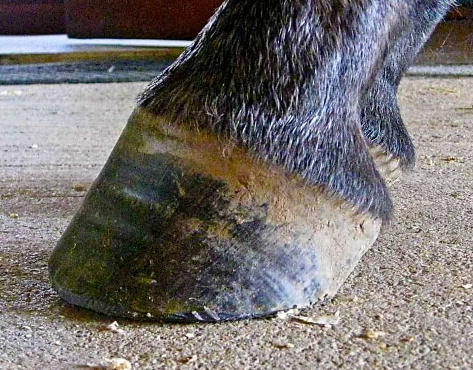


The Tolerable Club Foot The Horse
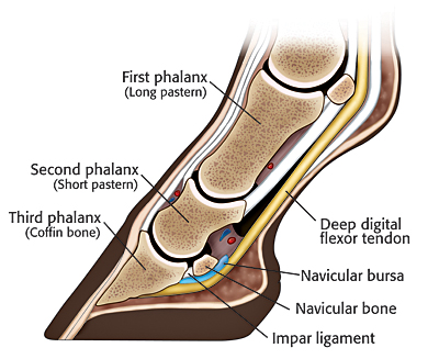
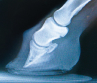
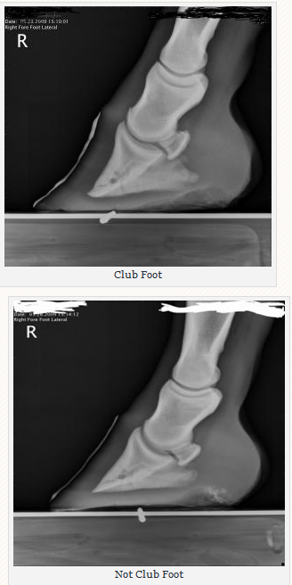
Radiograph shows a moderate flexural deformity (yellow circle) involving the DIPJ in a horse with a club foot The flexural deformity is caused by a shortened DDF muscle tendon unit (red line) Grossly, the dorsal hoof wall angle is upright or steep accompanied by aApparently the club foot condition has been with this horse since it was a foal whereas a dish at or just above the end of the toe would likely be considered grade 1 or 2 This club foot, as seen in photo 2, Photo 9 is the lateral xray showing the remodeled bone and poor quality of the boneIn this edition of the IMV Imaging Journal Club, Clinical Manager Laura Quiney seeks to answer the question What is new in equine foot radiography?' Webinar Using the Whole Horse Approach 3818 Sponsored By Sound Horse Technologies The term whole horse has been used by many different equine professionals to describe a host of ideas and
Digital radiography has become a standard in equine practice Of course, these advancements come at a cost to the owner On average, you can expect one digital image to set you back about $50 or so When you consider that a standard set of views of a foot is five images, and a hock and stifle are both four, it doesn't take long to do the mathCategories of club foot, on basis of joint motion and ability to reduce the deformities 11 i Soft foot also called postural foot can be treated by physiotherapy and standard casting treatment ii 2 Soft >Data publikacji paź 23, 14
Any foot radiograph for alignment First, you can evaluate the relationship of the tibia to the hindfoot, then the relationship of the hindfoot to the midfoot, and finally the relationship of the midfoot to the forefoot As you read the case scenarios, you will see how this can be aA lateral radiograph of the hoof is taken to confirm the position of the needle prior to injection Ideally the needle tip is midway between the proximal and distal borders Once the needle position is confirmed, the bursa is injected with the contrast mixture and a second lateral hoof radiograph is taken to confirm the filling of the bursaPedal osteitis is a common condition in the horse, resulting in ongoing foot pain and lameness, which can be extremely limiting to athletic performance The end result for many horse owners is rest, specific shoeing strategies, medications, and in some cases surgery is warranted Despite these treatment options, many horses do improve in their soundness,


Recognizing Various Grades Of The Club Foot Syndrome


Michael Porter Equine Veterinarian November 12
Seventyfour feet from 52 lame horses were included Twenty parameters were measured on radiographs, whereas the signal intensity, homogeneity and size of each structure in the foot were evaluated on magnetic resonance images The data were analysed using simple linear correlation analysis and classification and regression trees (CARTs) ResultsFoot laminitis 06 LM radiograph, illustration relating to horses including description, information, related content and more RossdalePartners Equis ISSN Related terms All information is peer reviewedIn this edition of the IMV Imaging Journal Club, Clinical Manager Laura Quiney seeks to answer the question What is new in equine foot radiography?'


Chapter 7 Unsoundness Of Horses Unsoundnesses Of The


Club Foot Or Upright Foot It S All About The Angles American Farriers Journal
A horse with a club foot is kind of like a horse in high heels The hoof angle becomes raised and the horse walks on his toe due to a shortening ofRadiography of the feet is commonly indicated for lameness diagnosis or followup, balance assessment in conjunction with a farrier, or for prepurchase examinations The purpose of this critically appraised topic was to identify recent scientific advances in foot radiography in order to direct the practice of evidencebased radiographyBy Christy West, TheHorsecom Webmaster Article # 9805 When you look at a radiograph (X ray) of a horse's foot, do you visualize soft tissues, or do you only see bones?


Recognizing Various Grades Of The Club Foot Syndrome
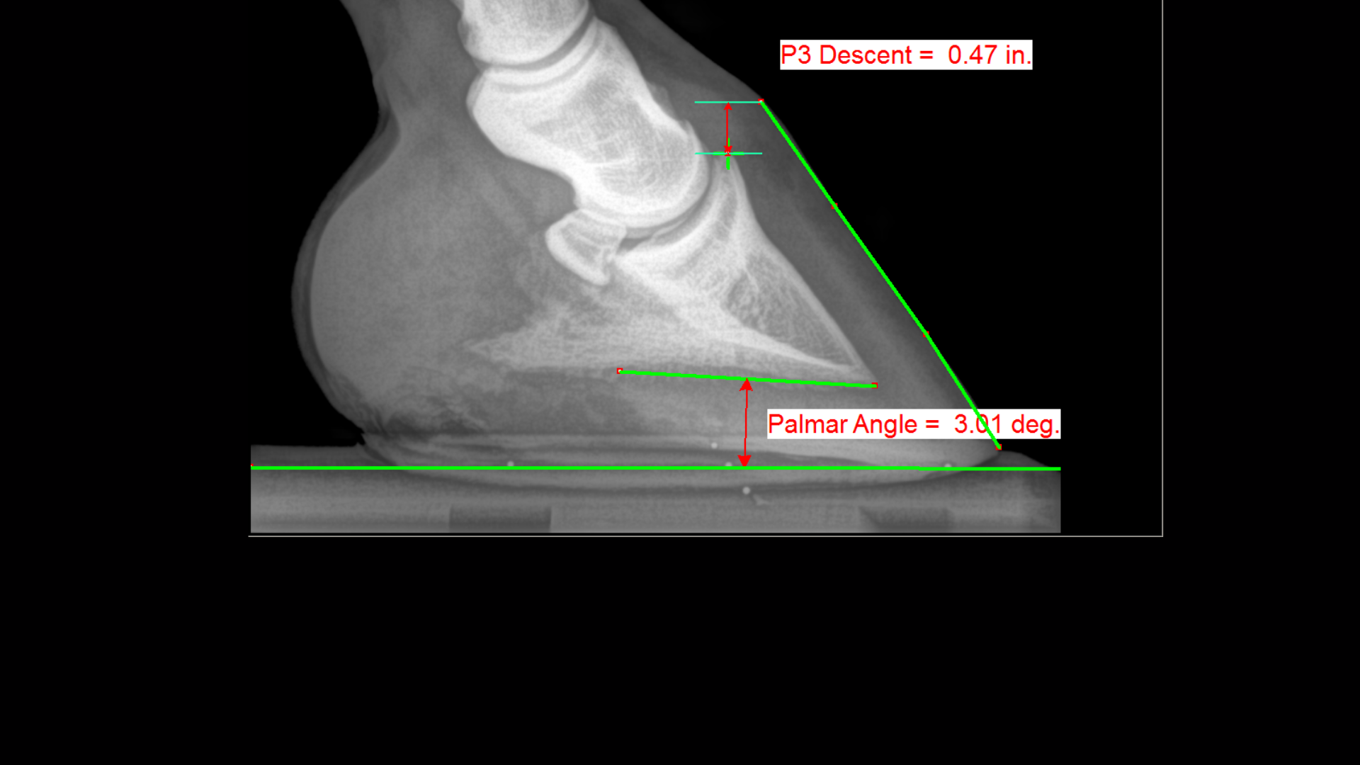


Podiatry The Equine Center
Caused by abnormal contraction of the deep digital flexor tendon, a club foot puts pressure on the coffin joint and initiates a change in a hoof's biomechanics Telltale signs of a club foot may include an excessively steep hoof angle, a distended coronary band, growth rings that are wider at the heels, contracted heels, and dished toesThe radiograph below is from a weanling colt with a severe case of a club foot Figure 1 is the affected foot and figure 2 is the normal foot Xray vision was not necessary in this case to confirm the diagnosis due to the classic distortion of the hoof capsuleExplore LISA's board equine clubfoot on See more ideas about horse health, equines, horse care


Natural Angle Volume 15 Issue 1 Spanish Lake Blacksmith


Hoof Radiographs They Give You X Ray Vision Part 3 Daisy Haven Farm
Foot is the standing dorsopalmar view In this view the horse is standing flat and the xray beam is centered halfway between the coronary band and the solar surface of the foot This generally requires that the horse be standing on a block this raises the footThe difference is readily determined by physical and radiographic examination The normal pedal bone angle for most horses is between 42 and 48 degrees, when the physical angle of the pedal bones are greater than 48 degrees and both feet present with a more 'boxy' shape than normal, Club Foot Syndrome can be confidently diagnosedIf your horse is lame, your vet probably decided what area to radiograph based on the results of an examination, and blocks can be especially helpful When your vet blocks your horse, she'll inject a local anesthetic substance into nerves supplying an area, or directly into a joint or other enclosed structure


1


Comstock Equine Hospital Veterinarian In City State Country Corrective Trimming
Symptoms of Club Foot in Horses Lameness Pain Excess toe wear Shortening of the tendon that is attached to the coffin bone Impacts the standing or movement of your young horse It can affect one or both limbs usually in the fore limbs Coronary band may bulge as the deformity progressesThe equine club foot is a tricky issue Radiograph showing a flexion contracture of the coffin joint with increased hoof angle, often associated with dishing of the dorsal hoof wallInequality in the angles of a pair of hooves (usually front), ie one hoof grows steeper than the otherRadiograph video sequencing of a horse with displacement of the coffin bone due to laminitis (Andrew van Eps Chris Pollitt)
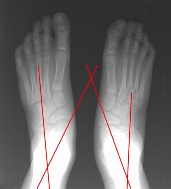


Clubfoot Imaging Practice Essentials Radiography Computed Tomography


The Chronicle Of The Horse
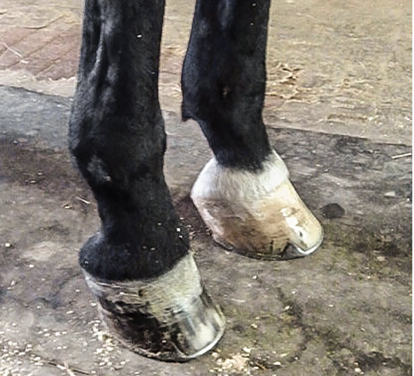
Almost always, I am called to club foot cases when the horse goes lame on the normal side This is usually caused by people trying to force the hooves to match each other This thinking often leads a farrier to cut the sole out from under P3 at the toe and allow the heels to grow unchecked on the normal foot Two wrongs don't make a right Treat the hooves as individuals, figure out theClub foot horses images A club foot can have significant repercussions on a horse's performance success and athletic longevity Prompt recognition and diligent farrier care allow the horse with a flexural deformity to Symptoms of Club Foot in Horses Lameness Pain Excess toe wear Shortening of the tendon that is attached to the coffin boneA horse with an upright alignment of the pastern bones will also have upright hoovesa situation that is sometimes mistaken for club foot A true club foot is significantly more upright than the other hooves, or the angles of both hoof walls are steeper than the angles of the pasterns The severity of the problem is commonly graded on a four


Epublishing


Managing The Club Hoof Easycare Hoof Boot News
Forward (club foot) or if the axis is broken back (long toe underrun heel), the radiograph will reveal the degree of deformity and the best way to trim the foot to improve it 1 Volume 6 Issue 2 A PUBLICATION OF PRACTICAL IDEAS AND SOLUTIONS FOR FARRIERS CONTINUED ON PAGE 2 RADIOGRAPHS FOR THE FARRIER Fig1An X ray of your horse's foot can help you predict the future while it shows you the present MECHANICAL LAMINITIS TREATMENT Foot X Rays A Crystal Ball?In the absence of lameness, detailed radiographic evaluation of the horse's foot is very useful in preplanning and management of trimming and shoeing of a horse's foot and in the prevention of lameness A horse's exhibiting poor foot conformation, imbalance, or abnormal patterns of growth can be clues to impending foot disease and lameness


Recognizing And Managing The Club Foot In Horses Horse Journals


What Advice Has Been Most Helpful When You First Encounter A Club Foot American Farriers Journal
A club foot is a DEFORMITY and for any horse to win at top level competition it needs every possible advantage and no drawbacks The only way to stop continuing problems with club footed horses is not to breed from them After 11 months of gestation, it is a costly and heart breaking exercise if it results in a club footed foal3 a recent method of classifying club feet using a grading system (grade 14) has been proposed 2,9 it would appear beneficial to classify the severity of theClubfoot, or talipes equinovarus, is a congenital deformity consisting of hindfoot equinus, hindfoot varus, and forefoot varusThe deformity was described as early as the time of Hippocrates The term talipes is derived from a contraction of the Latin words for ankle, talus, and foot, pesThe term refers to the gait of severely affected patients, who walked on their ankles


Surgical Services Scenic Rim Veterinary Service


Equine Podiatry Dr Stephen O Grady Veterinarians Farriers Books Articles
Any club foot that has been around a while will have a sensitive, unused, underdeveloped frog/digital cushion You can fix everything else and still have the back of the foot too sensitive for the horse to land on, which will cause the shortened stride and resulting club foot on its own – another vicious cycleThe search terms (horse* OR equine*) AND (foot OR feet OR digit* OR hoof OR hooves OR phalan * OR navicular) AND (radiograph* OR radiolog *) were generated and input into the PubMed search engine Following exclusion of studies more than 5 years old and those determined not to relate directly to the question, six useful results regardingClinical and Radiographic Examination of the Equine Foot 1 Introduction Lameness is one of the most frequently encountered problems in equine practice The foot


2


Handbook Of Equine Radiography Medicine Health Science Books Amazon Com
Or less and type 2 where the hoofground angle is greater than 90°And variable angle horizontal beam dorsopalmar foot radiographs were acquired while keeping the limb position constant Analyses of measurements demonstrated that hoof pastern angle had a linear relationship (R 2 = 0, P <Traditionally, club feet or flexural deformities have been classified as type 1 where the hoofground angle is 90°


2


Defining And Fixing A Horse S Club Foot American Farriers Journal
The lateral radiograph confirmed how little foot we really had to work with, which the horse's pain level and hoof inflammation was certainly telling us What was also very interesting was the DP radiograph, since it also indicated significant medial lateral hoof imbalances that we could help this horse with at the timeA horse with a club foot is kind of like a horse in high heels The hoof angle becomes raised and the horse walks on his toe due to a shortening ofClub foot is defined as a flexural deformity of the coffin joint and is a common problem in young, growing horses Characteristics of a club foot are a prominent or bulging coronary band, a veryDorsopalmar foot radiographs were acquired while varying the lateromedial stance;


Laminitis Signalment Treatment And Prevention Flying Changes


Equine Therapeutic Farriery Dr Stephen O Grady Veterinarians Farriers Books Articles
Szacowany czas czytania 3 minClub Foot Horse Radiograph It s a club no one wants to join but many americans these days find themselves automatically eligible for the bill of the month club yacht vivacity foot figure 87c 1 frontal radiograph of foot demonstrates alignment of the long axis of talus with the base of second metatarsal line a (17) Radiographic measurements ofInadequate sole depth will usually be accompanied by excessive toe length The xray will show whether the hoof pastern axis is parallel If the axis is broken forward (club foot) or if the axis is broken back (long toe underrun heel), the radiograph will reveal the degree of deformity and the best way to trim the foot to improve it
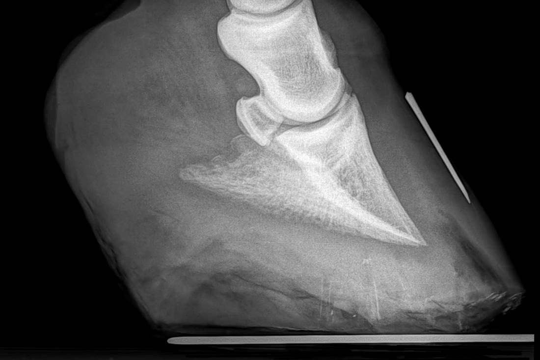


Understanding X Rays The Laminitis Site


Hoof Evaluation Radiographs For The Farrier
Szacowany czas czytania 8 minIn older horses, when club foot has become a more chronic condition, altering the biomechanics of the foot is often the key to management Radiographs of the feet are essential in providing the information necessary to evaluate the existing biomechanics and determining the appropriate changes that need to be madeThe contracted muscle/club foot condition is a common growth problem in young horses (up to 6 months of age), causing upright pasterns and a tiptoe stance This is often seen in foals with developmental problems due to rapid growth If discovered soon enough, this condition can be reversed by altering the foal's diet and reducing stress on


Shoeing Options For Club Foot In Horses


Michael Porter Equine Veterinarian 12
A horse with club foot has one hoof that grows more upright than the other The "up" foot is accompanied by a broken forward pastern, that is, the hoof is steeper than the pastern (Photo 1) In a normal foot, the hoof capsule and the pastern align Radiographs will show that the boneyLabels club foot, club foot radiograph, equine podiatry, Sammy L Pittman DVM Sunday, Setting the bar for success in my laminitis cases Welcome to 14!I wanted to review some of my laminitis cases that have proven very successful with regards to quickly adding sole mass and demonstrating an even hoof wall growth from toe to


Basic Shoeing Working With A Club Foot Farrier Product Distribution Blog


Basic Shoeing Working With A Club Foot Farrier Product Distribution Blog
Radiographic examination of the foot is one of the most frequently performed studies of equine limbs 2 It is somewhat more complex than other radiographic studies of horse limbs, but with proper equipment and technique excellent quality radiographs can be made Without a good quality radiographic examination of the foot, correct radiographic diagnoses cannot beClub Foot Etiology congenital Imaging CAVE – Cavus (high arched foot), forefoot Adductus, hindfoot Varus / Equinus — AP radiograph overlap of talus and calcaneus, varus angulation of forefoot (metatarsus adductus), inversion of foot — Lateral radiograph parallel alignment of talus and calcaneus and equinus (upward) position of


2
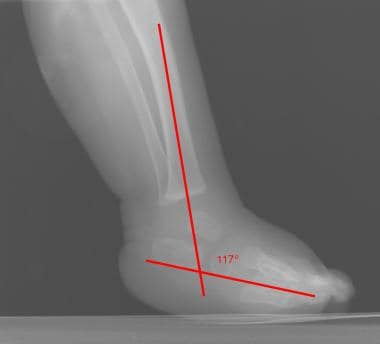


Clubfoot Imaging Practice Essentials Radiography Computed Tomography


Dr Rekha Ganeshalingam Club Foot Congenital Talipes Equinovarus Youtube


An Investigation Into The Association Between Plantar Distal Phalanx Angle And Hindlimb Lameness In A Uk Population Of Horses Clements Equine Veterinary Education Wiley Online Library


Understanding X Rays The Laminitis Site


2


Club Foot Horses Club Foot Feet Club


Farriery For The Hoof With A High Heel Or Club Foot Semantic Scholar


What S New In Equine Foot Radiology Imv Imaging


The Progressive Equine Services Hoof Care Centre Facebook


Understanding Club Foot The Horse Owner S Resource


Equine Podiatry Say What Mobile Veterinary Services


Boarding At Yucca Veterinary Medical Center


Recognizing Various Grades Of The Club Foot Syndrome


2
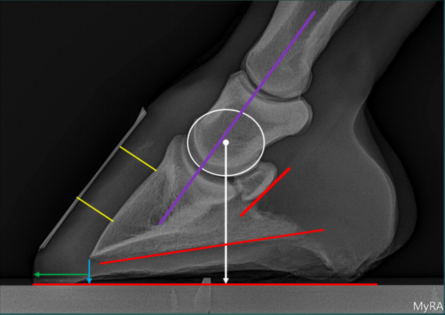


Podiatry Burwash Equine Services


Recognizing And Managing The Club Foot In Horses Horse Journals


Founder 13 Yo Paso Fino Barefoot Hoofcare


2


The Wild Mustang Hoof


Clubfoot Barefoot Hoofcare


Equine Podiatry Dr Stephen O Grady Veterinarians Farriers Books Articles


Recognizing And Managing The Club Foot In Horses Horse Journals


Recognizing And Managing The Club Foot In Horses Horse Journals


Radiolucency Glue On Horseshoes By Sound Horse Technologies


Pdf How To Perform Radiographic Guided Needle Placement Into The Collateral Ligaments Of The Distal Interphalangel Joint Semantic Scholar


2


Low Foot Case Study Dixie S Farrier Service


Recognizing And Managing The Club Foot In Horses Horse Journals
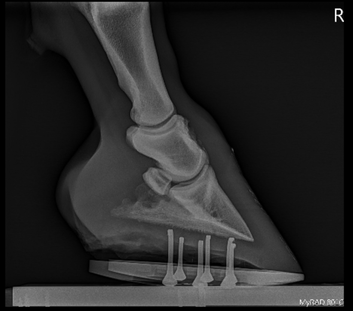


Podiatry Burwash Equine Services
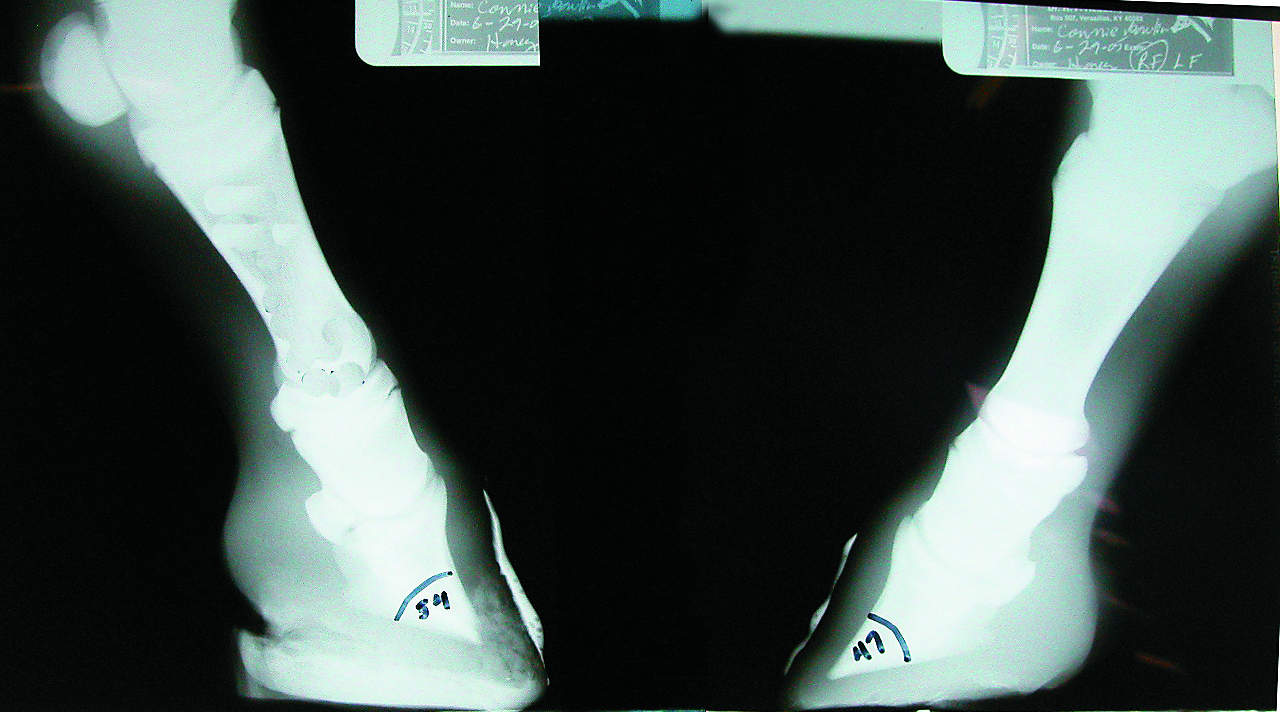


Foal With Hoof Problems Club Foot The Horse


2


2


Foal With A Club Foot Download Scientific Diagram
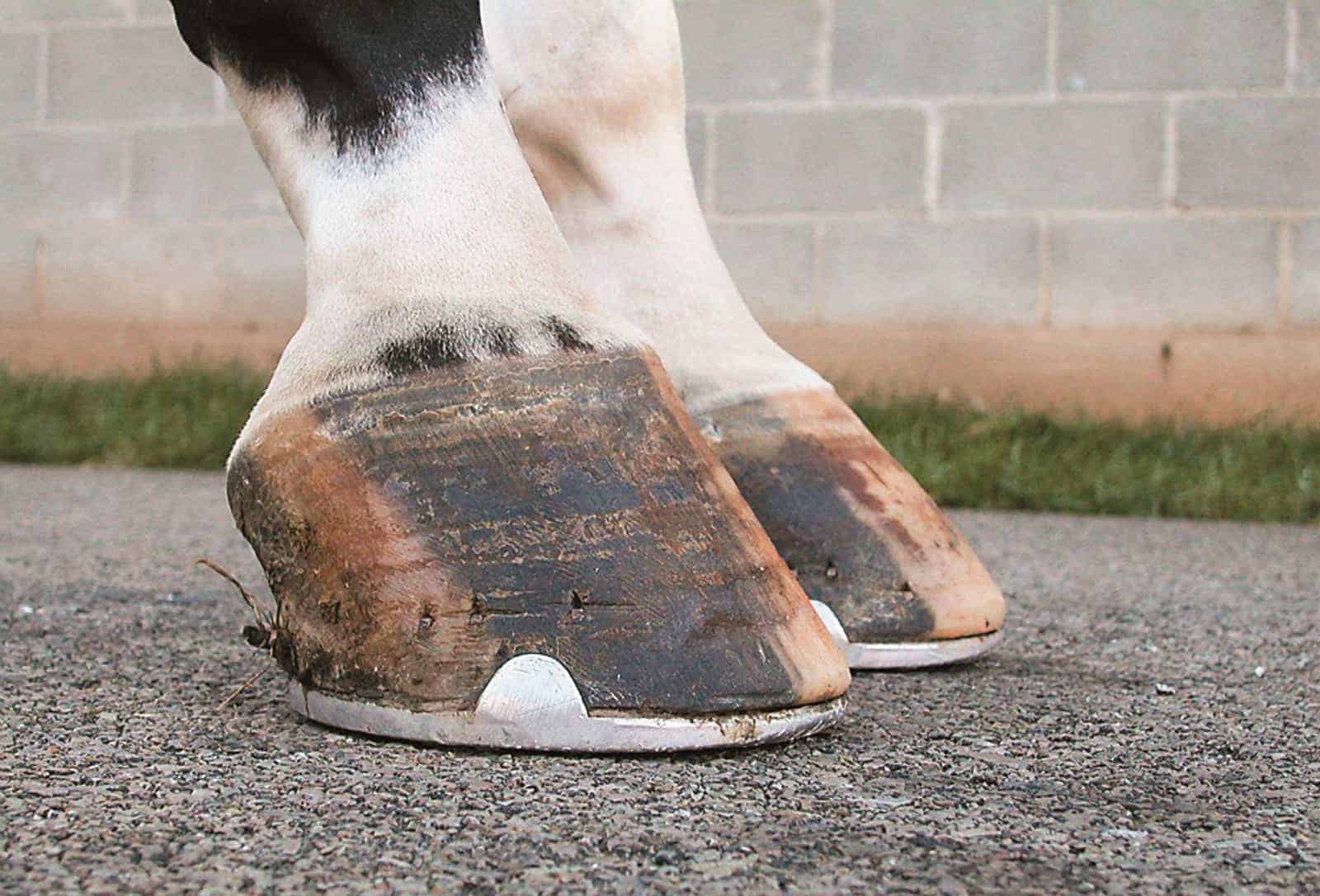


Managing The Club Foot The Horse


Hoof Conformation Vs Horse Conformation Scoot Boots Retail


2


Club Foot In Horses Brian S Burks Fox Run Equine Center Facebook


Thrush Cured Cowboy Pads And Copper Sulfate


Osteoarticular Radiographic Findings Of The Distal Forelimbs In Tbourida Horses Sciencedirect


Severe Lameness Scarsdale Vets
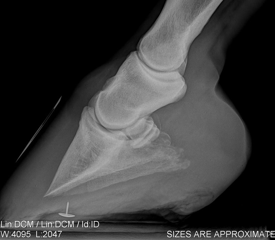


Understanding X Rays The Laminitis Site


Clubfoot Barefoot Hoofcare
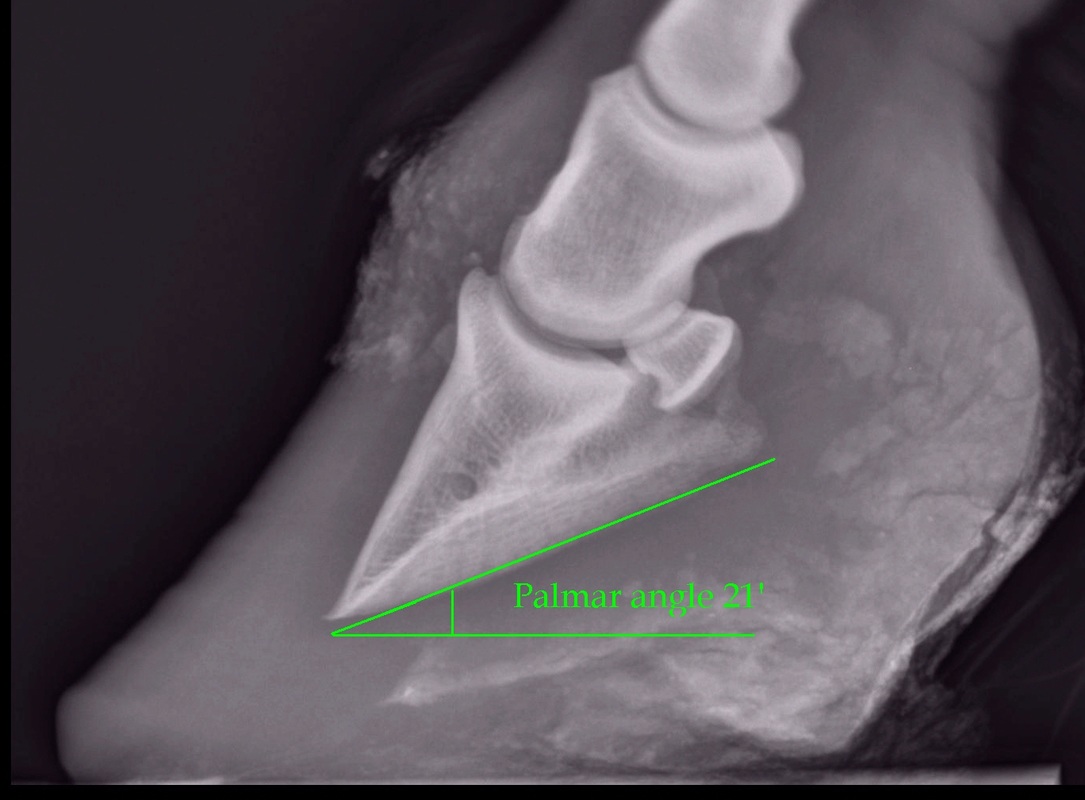


Understanding X Rays The Laminitis Site


The Wild Mustang Hoof


Low Foot Case Study Dixie S Farrier Service
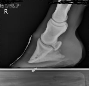


Clubfoot Barefoot Hoofcare


Lesson 4


Hoof Evaluation Radiographs For The Farrier
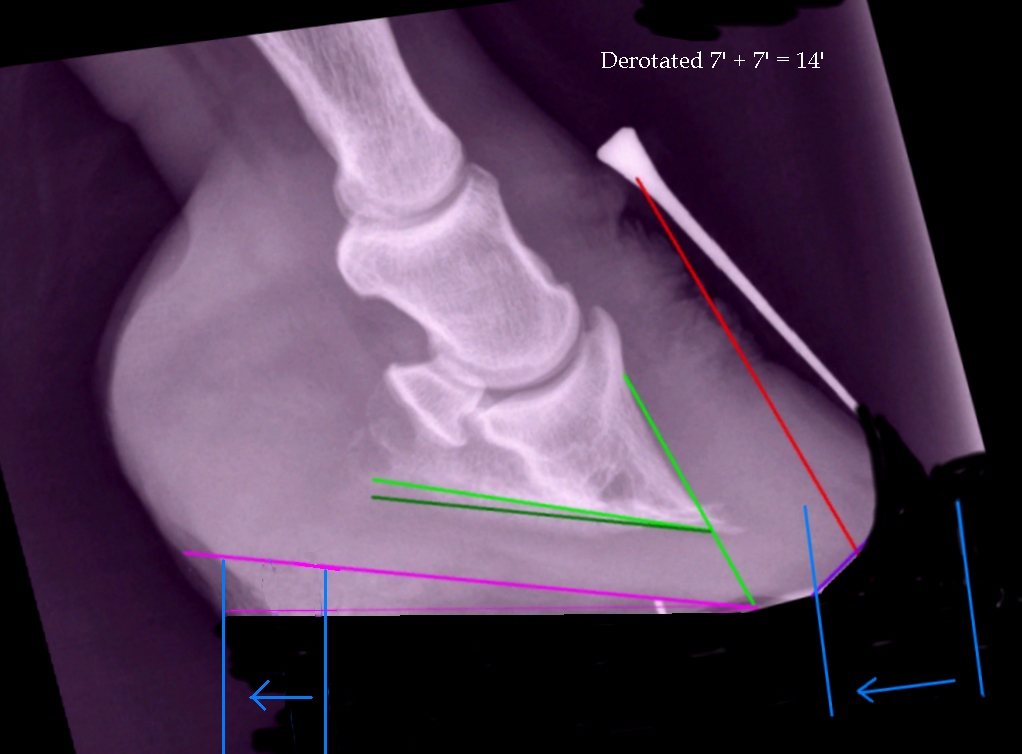


High Heels The Laminitis Site


Horse Club Foot


Clubfoot Barefoot Hoofcare
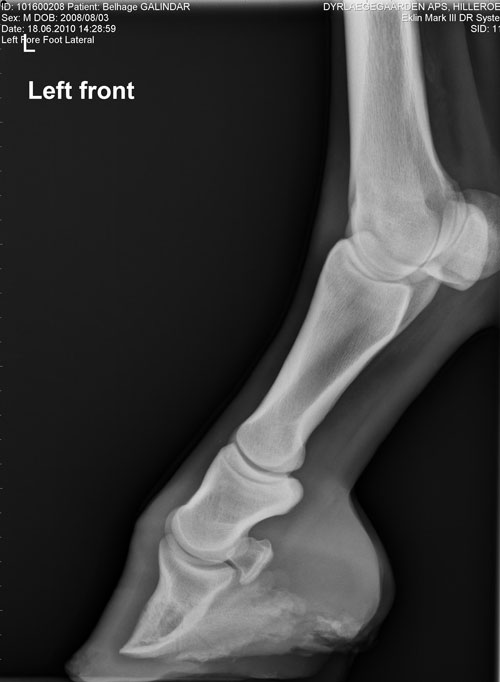


Contact Us Palmetto Equine Veterinary Services
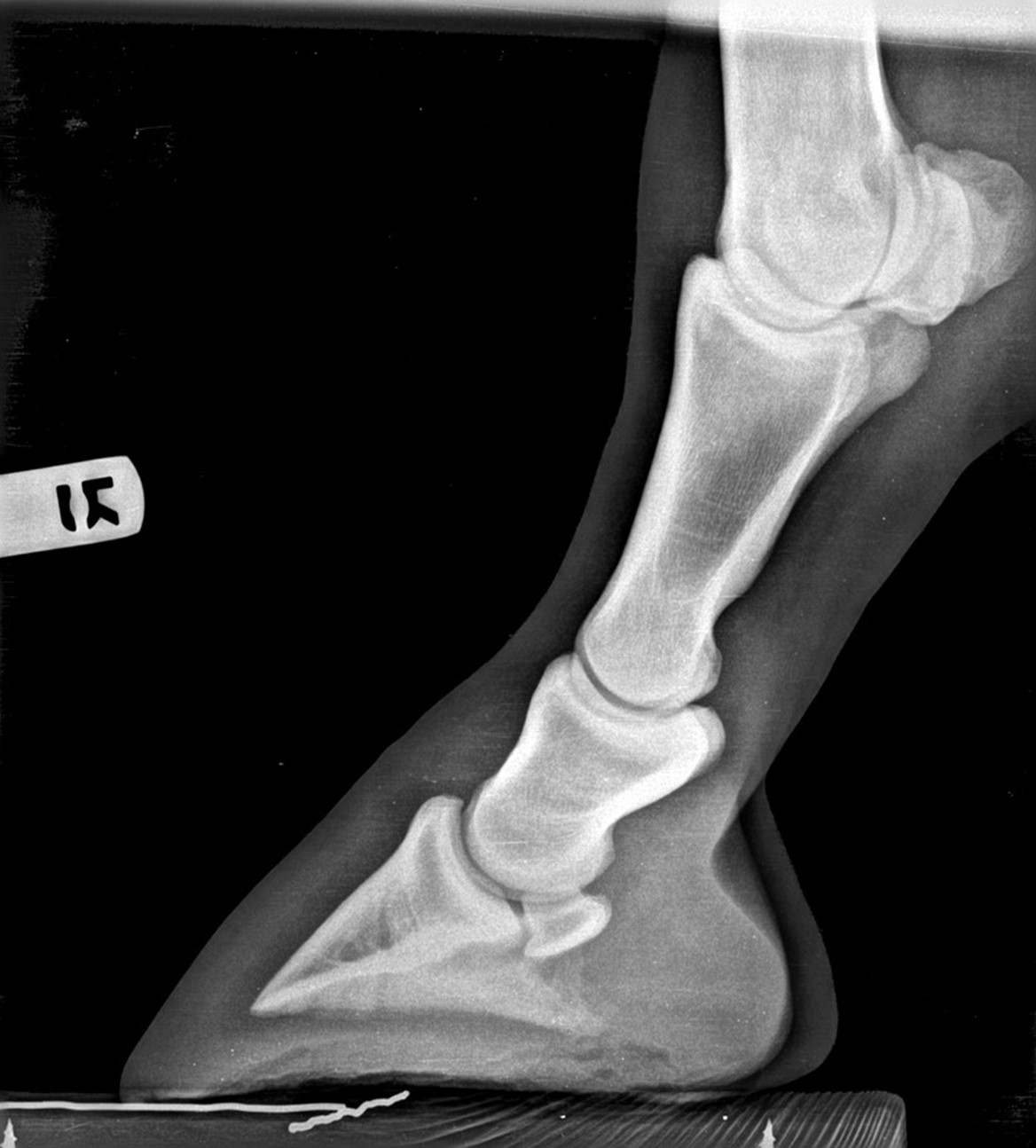


Hoof Radiographs Springhill Equine Veterinary Clinic


How To Treat Club Feet And Closely Related Deep Flexor Contraction


Hoof Evaluation Radiographs For The Farrier


The Tolerable Club Foot The Horse


New Strategies For Treating Club Foot From Foal To Adult


Hoof Angles Part 5 Enlightened Equine


2


Recognizing And Managing The Club Foot In Horses Horse Journals


Equine Therapeutic Farriery Dr Stephen O Grady Veterinarians Farriers Books Articles
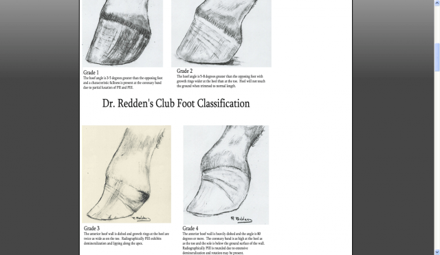


Managing The Club Hoof Easycare Hoof Boot News


2


Club Foot Horse Shefalitayal
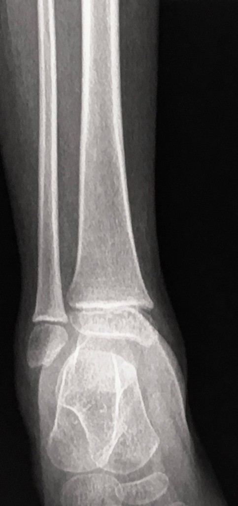


Equinus Foot Deformity Radiology Case Radiopaedia Org


Foal With A Club Foot Download Scientific Diagram
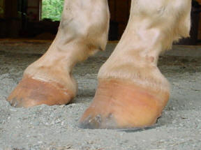


Club Foot


Farriers Archives Springhill Equine Veterinary Clinic


Equine Podiatry In Wendell Nc Neuse River Equine Hospital


The Wild Mustang Hoof


2


1


3


0 件のコメント:
コメントを投稿